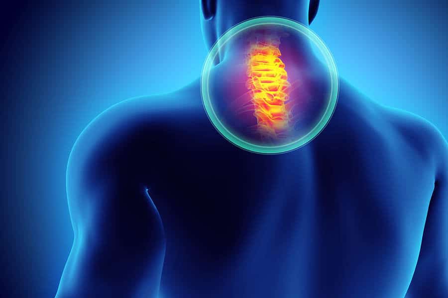
Human Spine Anatomy and Physiological
The human spine extends from the lower back to the brain, and is part of the central nervous system. It consists of a spinal cord, the outer layer of which is made up of myelin-sheathed nerve fibers. It has specialized tracts that send impulses such as pain, pressure and other stimuli. The cord is then covered and protected by vertebral column, which is also called spinal column. It has 33 bones (7in cervical region, 12 in thoracic region, 5 in the sacral region, 5 in the lumbar region, and another 4 in the coccygeal region).
I. Spinal Curves
The spine is naturally S-shaped, which is why it’s called spinal curve. Two regions (the cervical and lumbar) both have slight concave curve. The other two regions (the thoracic and sacral) have convex curve. The S-shaped curve helps the body maintain balance, absorb shock and allow a range of motion, by working like a coiled spring.
II. Vertebrae
This is the bones that interlock to protect the spinal cord and form the entire spinal column. It has 33 individual bones, all in all, divided in five regions: cervical, thoracic, lumbar, sacrum, and coccyx. Each region has unique feature and serve a distinct function. Of the 33 bones, only 24 are movable.
- Cervical (neck)
The cervical spine is otherwise known as the “neck.” Its main function is to support and balance the head. It has two specialized vertebrae connected to the skull, which makes the neck the most moveable part of the spine. It also has a peg-shaped axis which allows the neck to move sideways. Due to the wide range of movement, one is prone to neck pain. If you are suffering from such, check us out.
- Thoracic (mid back)
The thoracic is called the mid back. It holds rib cage that protects the lungs and heart. There are 12 thoracic vertebrae (T1 to T12). However, unlike the neck, its range of motion is limited.
- Lumbar (low back)
The lumbar is known to many as “low back.” Its main purpose is to support and hold the weight of the body. It has five vertebrae (L1 to L5). These vertebrae are larger and sturdier in order for them to absorb the shock when carrying and lifting heavy objects. Sometimes, people may suffer from Cervical Radiculopathy, which emanates from the neck and radiates to other parts of the body. If this happens to you, give us a call and we will alleviate the pain.
- Sacrum
This region connects the spine to the iliac or hip bones. There are five vertebrae in this region, which are all fused together. This and the hip bones form the pelvic girdle.
- Coccyx region
This is also called the tailbone. It has four fused bones. The coccyx attaches the ligaments and muscles to the pelvic floor.
III. Intervertebral discs
The intervertebral discs serve as a cushion to each of the vertebra. It separates each bone and keeps them from rubbing together. It has an outer ring that consists of criss-crossing fibrous brands. These pull the vertebral body from the elasticity of the nucleus. The inner part of the disc is the nucleus, which is a gel-filled center that is mostly composed of fluid. It allows the vertebral body to roll over the gel.
IV. Vertebral arch & spinal canal
These are bony projections found at the back of each vertebra. Each arch is composed of two laminae and two supporting pedicles. On other hand, the spinal canal is a hollow space that contains the cord, ligaments, fat and blood vessels.
Get Back Your Normal Life Again
As pain specialists, we can guarantee that we are more than qualified in alleviating your pain and treating your condition.
V. Facet joints
These are the joints in the spine that allow back motion. There are four facet joints in every vertebra. The two joints (superior facets) connect to the upper part of the vertebra, while the other two joints (interior facets) connect to the lower part.
VI. Muscles
There are two muscle groups in the spine: the extensors and flexors. The extensors, which are attached to the back part of the spine, allow people to lift objects and stand up. The flexors, which are found in the front, enable people to bend forward and flex. It also controls the arch in the lower back. These muscles help stabilize the spine.
VII. Ligaments
These are strong fibrous brands. They protect the discs, stabilize the spine and hold together the vertebrae. There are three ligaments found in the spine: the ligamentum flavum, the posterior longitudinal ligament (PLL), and the anterior longitudinal ligament (ALL). The ligamentum flavum attaches the lamina of every vertebra. The ALL and PLL prevent excessive movement in the vertebral bones; they run along the vertebral bodies, from the bottom to top of the spinal column.
VIII. Spinal cord
The spinal cord, which is generally 18 inches long, is found within the spinal canal, and runs from the brainstem down to the first lumbar vertebra. It ends there and separate into cauda equine, where it continues down to the tailbone. The main function of the spinal cord is relaying messages to and fro the brain and body. It is responsible for the sending of motor messages throughout the body from the brain. That’s why we feel and react. There are instances where one immediately reacts without it sending sensory messages to the brain. This is due to the spinal reflexes or special pathways. The nerve cells in the spinal cord are upper motor neurons, while the nerve cells down your back are lower motor neurons.
IX. Spinal nerves
There are 31 pairs of spinal nerves, which act as “telephone lines.” With their help, messages are being sent back and forth between the spinal cord and the body. It helps control movement and sensation. Every spinal nerve is composed of two roots: ventral and dorsal. The ventral is the front root that carries motor impulses from the brain, while the dorsal is a back root that carries impulses to the brain. The nerves run down the spinal canal, until it reaches the intervertebral foramen. From here, the nerves branch out into posterior primary ramus (smaller branch) and anterior primary ramus (larger branch). Each of these branches has sensory and motor fibers.
X. Coverings & spaces
Three membranes cover and protect the spinal cord: the pia mater (inner), the arachoid mater (middle), and the dura mater (outer). There are spaces found between each of these membranes. There are spaces between each of these membranes. These spaces are used in treatment procedures and diagnostics. The space between the inner and middle space is the subarachnoid space, while the space between the outer membrane and the bone is the epidural space.

